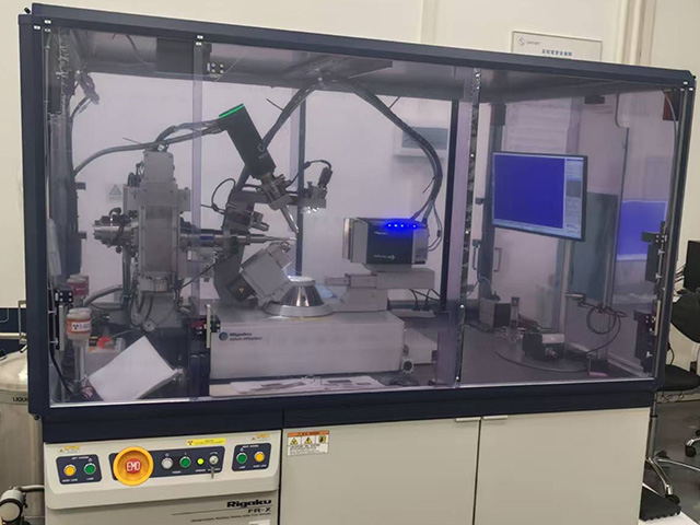
1.X-ray Source: Maximum rated output power 2.97kW
2. X-ray Source Maximum Operating Voltage: ≤45kV; Operating Current: ≤70mA
3. X-ray Source Target Focal Spot Size: No larger than 0.07mm × 0.07mm (point focus)
4. Anode Target Material: Cu, characteristic wavelength not shorter than 1.54 Å
Two-Dimensional Array Area Detector
(1) Detection Method: Direct counting of X-ray photons, not CCD or CPAD detection mode, no phosphor screen or similar for X-ray conversion required
(2) Arcing detection surface, capable of collecting data over a 2theta angle range of 150 degrees in a single exposure. Vertical height not less than 80mm
(3) Point Spread Function (FWHM): 1 Pixel
(4) Pixel Size: 100μm × 100μm
(5) Readout Dynamic Range: Up to 31 bit, each pixel contains two 16-bit photon counts
(6) Data Transmission Modes: 31bit, dual-channel 16bit, etc.
(7) Switching time between two photon counters in each pixel: ns level
(8) Readout Time: 7.4ms mode and 0ms mode
Goniometer (Kappa Goniometer)
(1) Kappa Axis Range: -177°~ +177° (collision-free conditions)
(2) Φ Axis Range: -360°~ +360°
(3) 2θ Axis Range: -82° ~ +95° (collision-free conditions)
(4) ω Axis Range: -176°~+176°
(5) Sample-to-Detector Distance: 45 ~ 150 mm (motorized step) (collision-free conditions)
(6) Centering Error: No greater than 7μm
Sample Cooling Device: Liquid nitrogen low-temperature blowing
(1) Temperature Range: 80-400K
(2) Temperature Stability: ±0.1K
Crystalline samples (single crystals of substances such as organic compounds, metal complexes, biological macromolecules, etc.) with a size preferably above 0.05 mm. The crystal surface should be clean, with consistent color and transparency. It should be free of attached small crystals, and free from defects such as cracks, overlaps, or imperfections.
Precise determination of the three-dimensional structure and electron density map of single crystals such as organic compounds, metal complexes, and biological macromolecules (proteins, nucleic acids, peptides, carbohydrates, etc.). This enables the acquisition of microscopic information regarding bond lengths, bond angles, torsion angles, molecular configurations, and conformations, and facilitates the study of their patterns, thereby further elucidating material properties and structure-function relationships.
 Return to Main Site
 Search
 中文
中文


 中文
中文


 Message