1.Solid-state lasers:
(405/445/488/515/561/638 nm).
2.Objectives:
2x apochromatic objective (N.A. 0.10, W.D. 8.5 mm)
4x apochromatic objective (N.A. 0.20, W.D. 20.0 mm)
10x apochromatic objective (N.A. 0.45, W.D. 4.0 mm)
20x apochromatic objective (N.A. 0.80, W.D. 0.8 mm)
40x apochromatic objective (N.A. 0.95, W.D. 0.21 mm)
60x apochromatic oil immersion objective (N.A. 1.42, W.D. 0.15 mm)
100x apochromatic oil immersion objective (N.A. 1.45, W.D. 0.13 mm)
40x apochromatic water immersion objective (N.A. 1.15, W.D. 0.63 mm)
3.Detectors:
Four GaAsP high-sensitivity detectors, with linear variable filters and spectral splitting; and one MA transmitted light detector.
4.NSPARC Detector:
Nikon's advanced spatial array detector with a maximum resolution of 8192 × 8192. 512 × 512 pixels at 30 fps; 1024 × 1024 pixels at 7.5 fps.
5.Standalone Camera:
Back-illuminated sCMOS sensor, maximum 5.5 megapixels.
6.Laser Confocal Scanning Head:
Galvanometer scanner: For high-resolution imaging, scan resolution of 8192 × 8192 pixels, scan speed: fast mode at 10 fps (512 × 512 pixels, bidirectional).
Resonant scanner: For high-speed imaging, maximum scan resolution of 2048 × 2048 pixels at 7.5 fps. Scan speeds: 2048 × 512 pixels at 30 fps, 2048 × 1024 pixels at 15 fps.
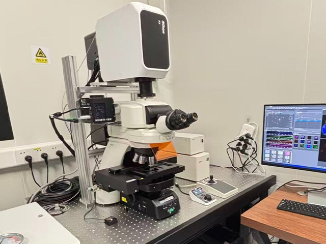
7.Fully Motorized Upright Microscope ECLIPSE Ni-E
8.Fluorescence Light Source: LED fluorescence light source, equipped with four LED sources (385 nm, 475 nm, 550 nm, and 621 nm) and red, green, blue fluorescence excitation blocks.
9.Equipped with a Live Cell Incubation System: Uses carbon dioxide gas as the source, ensuring long-term stability of temperature, humidity, and CO₂ concentration. Configured with adapters for 35 mm culture dishes, multi-well plates, and slides; compatible with the 40x water immersion objective.
Mainly used for confocal imaging of fixed tissue sections, cell culture slides, and live cells.
1.Equipped with four high-sensitivity GaAsP detectors, providing higher signal-to-noise ratio and clearer image contrast.
2.Equipped with a spatial array confocal NSPARC detector. Lateral resolution: 100 nm, axial resolution: 300 nm.
3.Equipped with a motorized stage for large-area image stitching, enabling high-resolution, large-field imaging.
4.Upright confocal high-speed resonant scanning mirror: simultaneously supports a scanning field of view (FOV) of 25 mm.
5.Equipped with an upright live cell workstation and a 40x water immersion objective for live cell imaging on an upright confocal microscope.
 Return to Main Site
 Search
 中文
中文


 中文
中文


 Message