1. Linear Measurement Range for Molecular or Nanoparticle Concentration:10 pM–100 nM.
2. Lower Detection Limit for Hydrodynamic Radius of Molecules or Nanoparticles:≤10 nm.
3. Dissociation Constant (K<sub>) Measurement Range for Molecular Interactions:pM–μM.
4. Single-Molecule Burst Analysis:Supports calculation of fluorescence resonance energy transfer (FRET) efficiency.
5. Signal Acquisition Optical Path:Integrated confocal optical path within the instrument; confocal volume ≤2 fL.
6. Laser Types:Continuous wave or picosecond pulsed lasers; laser types must include at least 488 nm, 561 nm, and 638 nm.
7. Laser Output Power:Continuously adjustable; maximum laser output power: ≥20 mW.
8. Sample Stage:X, Y, Z three-axis automated positioning sample stage; enables batch detection of 18 samples; sample chamber automatically measures and records experimental temperature.
9. Temperature Control System:Equipped with three temperature probes for real-time temperature measurement (sample chamber body, sample chamber lid, and sample);control precision of ±0.3°C under room temperature fluctuation range of ±1°C; control range: room temperature +3°C to 50°C.
10. Correlation Data Acquisition Card:Capable of real-time output of autocorrelation data from two channels and cross-correlation data from one channel; minimum correlation time <1000 ms.
11. Data Acquisition Software:One-click fully automated confocal pinhole calibration; customizable settings for data acquisition time and repetition次数; one-click start/stop; supports real-time display of fluorescence intensity signals, fluorescence autocorrelation, and cross-correlation decay curves.
12. Data Analysis Software:Built-in multiple analysis models available, including three-dimensional free diffusion model for molecules, fluorescence triplet state analysis model, and anomalous diffusion model. Supports detection and calculation of molecular concentration, hydrodynamic radius, and diffusion coefficient. Automatic or manual setting of fitting initial values; one-click data fitting.
Sample Preparation Notes:
1. Sample Quality and Labeling:
Prepare fresh, high-purity samples whenever possible. Except for autofluorescent samples (such as AIE materials, quantum dots, etc.), samples must be chemically labeled (amino labeling recommended) or fused with fluorescent proteins, and must be excitable by the 488/561/638 nm lasers equipped on the DEMO instrument. Additionally, the Stokes shift of excited samples should not be excessively large (must fall within the filter detection range of the corresponding detectors). Prepare the corresponding dye used for sample labeling as a separate control (e.g., if the sample is labeled with Alexa 647, prepare free Alexa 647 dye); recommended concentration is 1–100 nM, with a volume of 50–100 μL.
2. Sample and Buffer Preparation:
Prepare appropriate amounts of samples and buffers according to experimental objectives; aliquoting is advisable. It is best to prepare both high-concentration and low-concentration samples simultaneously.
3. Sample Types and Loading Volume:
Homogeneous solutions and cell lysate samples can be used, but the medium must be aqueous. Live cell samples or samples with high organic solvent content (>50%) are not permitted. Generally, the loading volume for a single FCS experiment should be ≥10 μL. If the acquisition time is long, the loading volume should be increased to avoid effects from sample evaporation; therefore, a loading volume of 30–100 μL is generally recommended. Furthermore, the optimal loading concentration for FCS experiments is 1–10 nM. If the sample has low photon quantum efficiency, the loading concentration may be increased; conversely, decrease the loading concentration.
4. Buffer Requirements:
The background signal of various buffers used in the experiment should not be excessively high—the cleaner, the better. Use 0.1 μm or 0.22 μm filters for filtration if conditions permit. The following conditions must also be met:
① Do not use contaminated or easily contaminated buffers; commercial buffers are preferred.
② Avoid adding surfactants (such as Triton X-100, etc.) to buffers whenever possible. If unavoidable, control the dosage; the amount should not exceed 0.5% of the total reaction system.
③ Avoid using reagents that affect solution viscosity, such as glycerol.
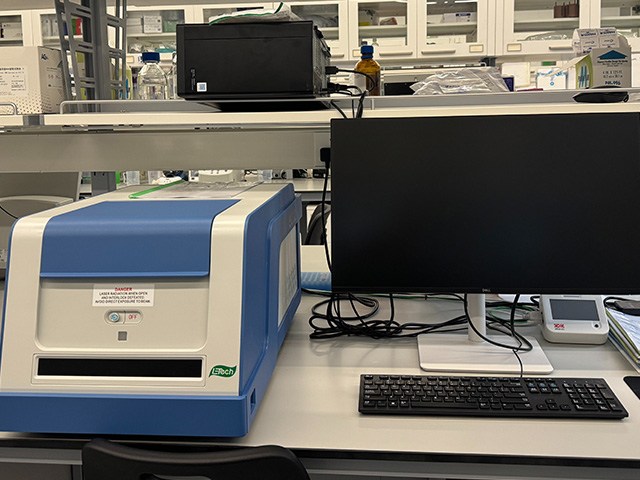
Utilizes fluorescence correlation spectroscopy (FCS) and fluorescence cross-correlation spectroscopy (FCCS) techniques to quantitatively detect the concentration, molecular size, intermolecular interactions, and conformational changes of molecules or nanoparticles in solution samples or cell lysates.
 Return to Main Site
 Search
 中文
中文


 中文
中文


 Message