The Biomedical Imaging Core Facility focuses on the construction of instrumentation and equipment to meet the scientific research needs for imaging spatial resolution, temporal resolution, and depth. It is supplemented by image processing workstations for multidimensional analysis, revealing the structure and function of organisms across multiple dimensions, from molecules, cells, and tissues to live small animals.
The Biomedical Imaging Core Facility is currently equipped with 19 large-scale instruments and 3 sets of professional image analysis software. The main equipment includes a super-resolution microscopy system (comprising 6 units, including STED-Facility Line, Multi-SIM, AXR-NSPARC, and LSM990), a laser confocal fluorescence microscopy system (3 units, including STELLARIS 8-FLIM, Evident Spin SR, and FV4000), a light sheet microscopy imaging system (LS18), a wide-field scanning imaging system (3 units, including the VS200 slide scanner, ECLIPSE Ti2-E line-scanning Raman microscope, and ECLIPSE Ji multimodal imager), a small animal live imaging system (4 units, including FV4000MPE and Insight-RSPAM3), a biological imaging sample preparation system (TCS-23), and a spatial molecular imaging system (CosMx Space Molecular Imager).
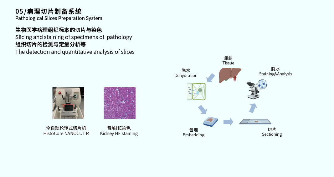
In terms of spatial resolution, the facility incorporates a super-resolution confocal microscope (STED-Facility Line) with an XY resolution of up to 30 nm, ensuring clear capture of fine structures within organisms. For temporal resolution, the full-frame frame rate can reach up to 89.1 fps (Evident Spin SR), and it is equipped with a live-cell workstation, enabling real-time tracking of dynamic biological processes as well as continuous, long-duration imaging. For depth detection, the facility is equipped with a light sheet microscope (LS18), a two-photon microscope (FV4000MPE), and a laser scanning photoacoustic microscope (Insight-RSPAM3), enabling precise imaging of deep tissues (with a maximum detection depth of up to 10 mm). Combined with professional image analysis software, it comprehensively reveals the complex structures and functions of organisms across multiple dimensions, from molecules, cells, and tissues to live small animals. Additionally, the facility is equipped with a full-process pathological section preparation system, providing key technical support for the preparation of biological imaging samples.
The professional image analysis software configured in this facility includes 2 sets of Imaris multidimensional image analysis software and 1 set of Amira 3D for Cell Biology multidimensional image analysis software.
A total of 15 full-process technical capabilities have been established, as listed below:
Full-process Service Items of the Biomedical Imaging Core Facility/No. | Service Item | Involved Major Equipment | Affiliated Facility |
1 | Tissue Clearing, 3D Imaging, and Data Analysis Service | 1.Tissue Clearing Devices 2.Light sheet Microscope 3.Imaris & Amira | Biomedical Imaging Core Facility |
2 | Paraffin Tissue Dehydration and Embedding Service |
| Biomedical Imaging Core Facility |
3 | Paraffin Sectioning Service |
| Biomedical Imaging Core Facility |
4 | Hematoxylin and Eosin (H&E) Staining Service |
| Biomedical Imaging Core Facility |
5 | AB-PAS Staining Service |
| Biomedical Imaging Core Facility |
6 | Masson's Trichrome Staining Service |
| Biomedical Imaging Core Facility |
7 | Immunohistochemistry (IHC) Single-label and Dual-color Staining Service |
| Biomedical Imaging Core Facility |
8 | Full-process Service for Routine Pathological Tissue Dehydration, Embedding, Sectioning, and H&E Staining |
| Biomedical Imaging Core Facility |
9 | Live-cell Label-free Imaging | Live-cell Label-free Superesolution Confocal Microscope AXR-NSPARC/MH-SpotyView | Biomedical Imaging Core Facility |
10 | Confocal Laser Scanning Imaging | Upright Laser Scanning Confocal Microscopy AXR-NSPARC | Biomedical Imaging Core Facility |
11 | Super-resolution Imaging | Multi-mode Structured Illumination Microscope Multi-SIM | Biomedical Imaging Core Facility |
12 | Tissue Clearing Sample Preparation | Tissue Clearing Devices | Biomedical Imaging Core Facility |
13 | Light Sheet Imaging | Light sheet Microscope | Biomedical Imaging Core Facility |
14 | Confocal Laser Scanning Imaging | 1.Superresolution Scanning Confocal Microscope AXR-NSPARC 2.Confocal Laser Scanning Microscope FV4000 | Biomedical Imaging Core Facility |
15 | STED Imaging with Super-resolution Confocal Microscope | STED-Facility Line | Biomedical Imaging Core Facility |
Since its establishment, the facility has served approximately 60 research groups from both within and outside the institution, led the signing of six external service agreements, and provided external services to numerous universities, hospitals, and biomedical companies.
Recent key developments of the facility include: 1) Establishing comprehensive tissue clearing techniques, integrating tissue clearing, light sheet three-dimensional imaging, and image analysis into a closed-loop solution covering the entire process, thereby significantly improving result reproducibility through standardized workflows; 2) Optimizing multicolor fluorescence imaging acquisition workflows to address challenges such as crosstalk caused by spectral overlap and rapid photobleaching of fluorescence signals; 3) Developing live-cell label-free imaging technology to overcome the contradiction between traditional super-resolution imaging techniques and the requirements for long-term, high-fidelity live-cell imaging, enabling non-invasive high-resolution dynamic observation of living specimens; 4) Exploring spatial molecular imaging technology to achieve ultra-high and ultra-multiplex target detection at subcellular resolution, precisely resolve cellular boundaries, and obtain high-quality molecular information.
 Return to Main Site
 Search
 中文
中文


 中文
中文
 Home
Home

 18612629622
 yuanlan@smart.org.cn
 深圳市光明区卫光生命科学园1A栋二层

 Message