Welcome to SMART Core Facility
Welcome to SMART Core Facility
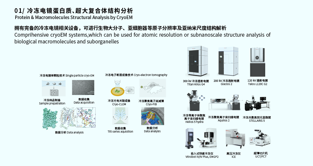
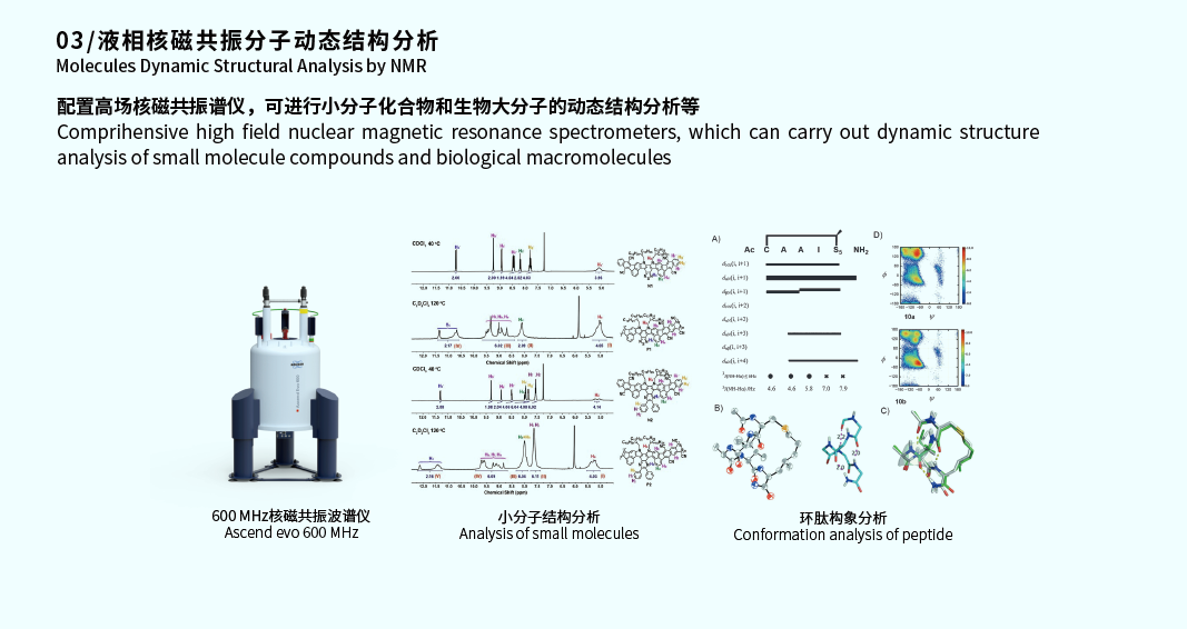
The Structural Biology Core Facility provides comprehensive support for structural biology research across biological molecules, subcellular structures, and cells through multi-scale structural analysis technologies. The facility integrates X-ray crystallography, nuclear magnetic resonance (NMR) spectroscopy, cryo-EM single-particle analysis, and cryo-electron tomography (Cryo-ET), delivering structural information from atomic to nanometer scales for diverse biological samples.
The facility is equipped with 20 state-of-the-art large-scale instruments, covering the entire workflow from sample preparation to high-resolution imaging and 3D reconstruction. Core instruments include two 300 kV cryo-TEMs (Thermo Scientific Krios G4) equipped with Falcon 4i direct electron detectors and SelectrisX energy filters, enabling stable data collection for single-particle analysis and cryo-electron tomography, supporting atomic resolution (typically better than 3 Å) structure determination. One 200 kV cryo-TEM (Glacios 2) is used for rapid cryo-grid screening and high-resolution imaging of nanoparticles, exosomes, membrane vesicles, and other biological macromolecules. Additionally, a 120 kV TEM supports negative staining and room-temperature ultrathin section imaging for rapid morphological assessment.
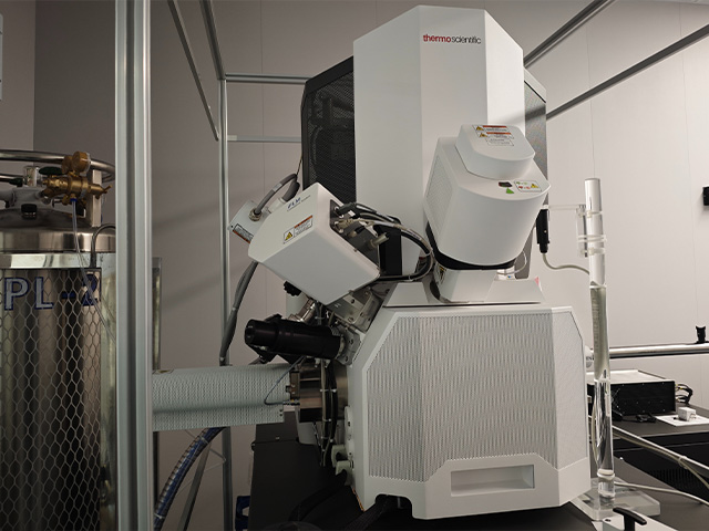
For in situ structural analysis, the facility features two cryo-focused ion beam scanning electron microscopes (Aquilos 2) and one cryo-plasma focused ion beam scanning electron microscope (Helios 5 Hydra CX), specifically designed for preparing thin lamellae (100–150 nm thick) from large-volume biological samples such as cells, tissues, and organs, providing optimal samples for cryo-electron tomography. Complementary cryo-sample preparation systems include plunge freezers, high-pressure freezers, and plasma cleaners, ensuring vitrification of samples under near-physiological conditions.
In data processing and analysis, the facility utilizes Amira 3D visualization software, supporting automated segmentation, 3D rendering, and video generation of electron tomography data, facilitating the visualization of complex biological structures.
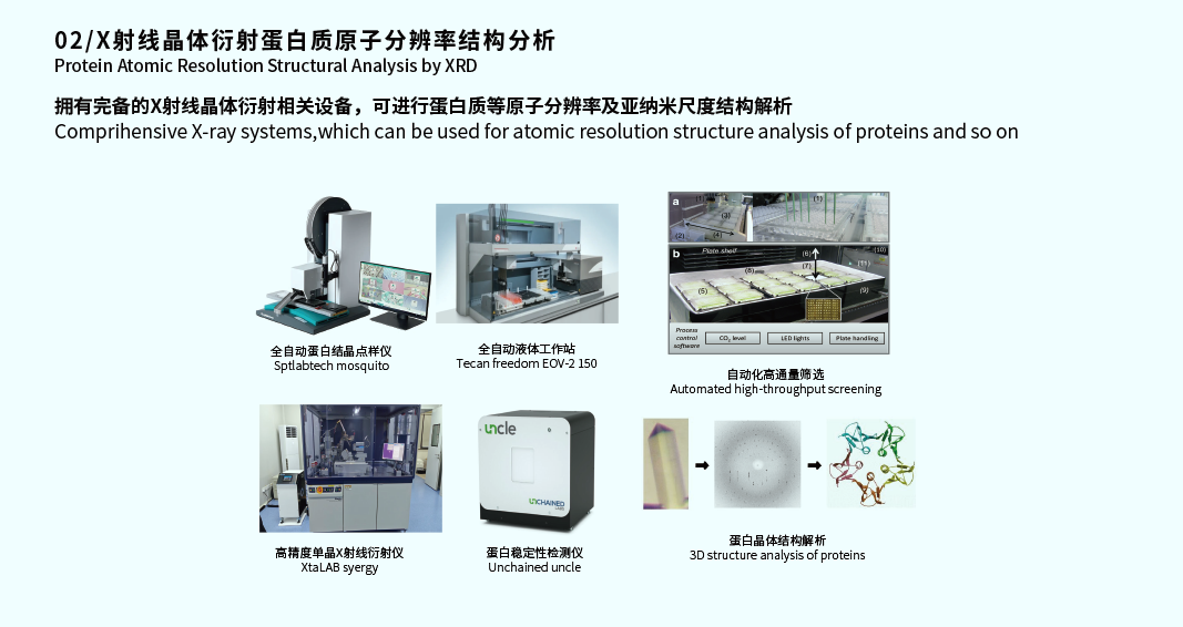
Beyond cryo-EM technologies, the facility also integrates a high-brilliance X-ray single-crystal diffractometer, a protein stability analyzer, and an automated liquid handling workstation, enabling full-process support from crystal screening and diffraction data collection to structure determination. This further expands the facility's multi-dimensional analytical capabilities in structural biology and drug discovery.
The facility has established six full-process technical services, listed below (Table 5.1).
No. | Service Name | Major Instruments Involved | Core Facility |
1 | Rapid Plunge-Freezing Sample Preparation | Plunge Freezer | Structural Biology Core Facility |
2 | Cryo-TEM Analysis (Single-Particle Samples) | Cryo-TEM | Structural Biology Core Facility |
3 | Cryo-TEM Analysis (Electron Tomography Samples) | Cryo-TEM | Structural Biology Core Facility |
4 | Characterization of Nanoparticles, Exosomes, Vesicles, and Nanocrystals | Cryo-TEM | Structural Biology Core Facility |
5 | Room-Temperature TEM Analysis | Room-Temperature TEM | Structural Biology Core Facility |
6 | One-Stop X-ray Diffraction Structure Determination | High-brilliance Single-Crystal X-ray Diffractometer | Structural Biology Core Facility |
Since becoming operational, the facility has provided technical support to 33 research groups, trained over 200 users, and facilitated breakthroughs in numerous scientific projects. The facility offers comprehensive technical services, including:
1) Rapid Plunge-Freezing Sample Preparation
This service aims to rapidly freeze biological samples into vitreous ice, preserving their native hydrated state and fine structure while preventing ice crystal damage. Using plunge freezers such as the Vitrobot Mark IV or EM GP2, liquid samples like purified protein complexes, viruses, or subcellular components are quantitatively applied to specific grids (e.g., copper or gold grids). By controlling environmental temperature and humidity, the plunge freezer rapidly immerses the grid into liquid ethane cooled by liquid nitrogen. This process (typically completed within milliseconds) solidifies the water molecules without crystallization, providing high-quality pristine samples for subsequent high-resolution cryo-EM observation. This technique is fundamental and essential for cryo-EM analysis, ensuring the observed biomolecules remain in a near-physiological state.
2) Cryo-TEM Analysis (Single-Particle Samples)
This service utilizes the 300 kV cryo-TEM (Thermo Scientific Krios G4) for high-resolution data acquisition of vitrified samples. The technical workflow includes:
High-throughput Data Collection: Utilizing the Falcon 4i direct electron detector and SelectrisX energy filter, automated, high-throughput image acquisition is performed on biological macromolecules uniformly distributed across the grid holes. The energy filter significantly enhances image contrast and signal-to-noise ratio by removing inelastically scattered electrons.
Atomic Resolution Structure Determination: Tens of thousands to millions of 2D projection images containing particles in various orientations are processed through complex image processing algorithms for particle picking, 2D classification, and 3D reconstruction, ultimately yielding a density map.
Model Building: Based on high-resolution density maps (typically better than 3 Å, reaching atomic resolution), facility technical staff assist users in building and refining atomic models of biological macromolecules, precisely revealing the 3D spatial structure, domain conformations, and interacting molecular mechanisms of proteins and their complexes.
3) Cryo-TEM Analysis (Electron Tomography Samples)
This service focuses on in situ structural biology, providing 3D structural analysis of cells, tissues, or macromolecular complexes. Leveraging the 300 kV cryo-TEM, the facility offers Cryo-electron Tomography (Cryo-ET) services:
Tilt Series Data Collection: For cryo-lamellae (typically 100–300 nm thick) prepared by cryo-FIB/SEM, a series of images is acquired as the stage is tilted over a range of angles (e.g., from -60° to +60°), generating a set of projection images known as a tilt series.
3D Reconstruction and Tomogram Generation: Software is used to align the tilt series images and perform 3D reconstruction, generating tomograms rich in structural information.
Visualization and Sub-tomogram Averaging: Tomograms contain the complex environment within cells, including the cytoskeleton, organelle interactions, and in situ protein complexes. Using 3D visualization software like Amira, facility staff can perform automated or manual 3D segmentation and rendering of target structures. Sub-tomogram averaging is applied to resolve high-resolution structures of in situ protein complexes with multiple copies, enabling understanding of the true arrangement and functional state of macromolecules within cells at near-atomic resolution.
4) Characterization of Nanoparticles, Exosomes, Vesicles, and Nanocrystals
This service targets smaller biological nanomaterials, primarily utilizing the 200 kV cryo-TEM (Glacios 2) or the 300 kV TEM for rapid, high-resolution morphological characterization:
Rapid Screening and Imaging: For samples like lipid nanoparticles, exosomes, and membrane vesicles, the 200 kV TEM provides an efficient quality screening pathway. While atomic resolution is not typically required, key features such as particle size, size distribution, morphological integrity, and multi-lamellar structure need clear observation.
High-Contrast Imaging: Cryo-TEM techniques allow direct observation of these samples in their native, solution-state environment, avoiding artifacts or deformations potentially introduced by traditional negative staining. For nanocrystals, this technique allows direct visualization of lattice fringes, morphology, and potential aggregation states, providing direct structural evidence for drug delivery systems, exosome biology, and nanomaterial science.
5) Room-Temperature TEM Analysis
This service is primarily for biological samples that do not require cryo-conditions, utilizing a 120 kV room-temperature TEM (Talos L120C) for rapid morphological assessment. Applications include:
Negative Stained Sample Observation: Purified protein complexes, virus particles, or organelles are treated with negative stain, where heavy metal salts outline the sample's morphology and contours. This is an effective method for rapidly assessing sample purity, homogeneity, and preliminary structure, often used for quality control screening before cryo-EM preparation.
Room-Temperature Ultrathin Section Observation: Observation of tissue or cell samples that have undergone chemical fixation, resin embedding, and sectioning with a room-temperature ultramicrotome. This is primarily used in cell biology research to observe subcellular ultrastructure, such as mitochondrial and endoplasmic reticulum morphology, cell junctions, and pathological changes in cells.
6) One-Stop X-ray Diffraction Structure Determination
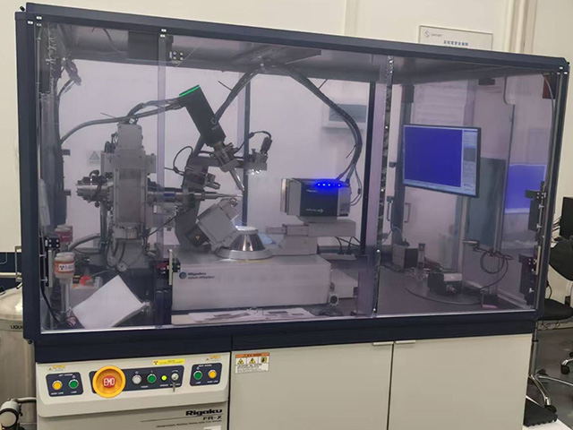
Relying on the high-brilliance single-crystal X-ray diffractometer (Rigaku Synergy FRX), the facility provides full-process services from crystal to atomic structure:
Crystal Screening and Optimization: Using an automated liquid handling workstation and a protein stability analyzer, the facility assists users in protein crystallization condition screening and optimization. The protein stability analyzer assesses protein monodispersity and thermal stability in different solution environments, providing guidance for crystallization screening.
Diffraction Data Collection and Processing: Well-diffracting protein crystals are mounted, and high-resolution diffraction data is collected using the high-brilliance X-ray source.
Structure Determination and Refinement: Using specialized software suites, diffraction data is indexed, integrated, and scaled. Phases are determined using molecular replacement or heavy atom methods, ultimately leading to the building and refinement of an atomic-resolution 3D protein structure. This technique complements cryo-EM and is particularly suitable for proteins or small molecule drug targets that crystallize readily and require precise atomic-level conformational detail.
In summary, these six services constitute a comprehensive, integrated structural biology platform, spanning from nanoparticle characterization to atomic model building, and from purified in vitro samples to in situ cellular structures.
Dr. Gang Fu received his Ph.D. from Hokkaido University and has worked on structural biology researches of macromolecular complexes at UT Southwestern Medical Center, Fudan University, and the University of Massachusetts Chan Medical School. He has mastered advanced electron microscopy techniques including room-temperature ultrathin sectioning, cryo-EM single-particle analysis, and cryo-electron tomography. The Structural Biology Core Facility integrates room-temperature and cryo-electron microscopes, X-ray diffractometer, NMR spectrometer, and supporting equipment to provide high-resolution imaging and in-situ structural analysis across multiple scales, from small molecules and protein complexes to organelles.
 18116093956
 fugang@smart.org.cn
 深圳市光明区卫光生命科学园5栋一层
Leiqing Yang obtained her doctoral degree from University of Science and Technology of China (USTC) in 2021.From2021-2023,Leiqing trained as postdoctoral fellow with Prof. Guoqiang Bi at Shenzhen Institutes of Advanced Technology (SIAT).She does research in the molecular and structural base of synapse formation using cryo-ET. Currently,she is mainly engaged in the operation and maintenance of cryo-FIB and cryo-CLEM. She has undertaken one regional joint youth fund project in Guangdong Province as a project leader, and participated in many National Natural Science Foundation of China and Brain Program youth projects. Her researches have been published in Neuroscience Bulletin, Frontiers in Cellular Neuroscience, FASEB Journal and other SCI journals.
15255151084
yangleiqing@smart.org.cn
深圳市光明区卫光生命科学园5栋一层
Dr. Lu graduated from the University of Macau and subsequently conducted postdoctoral research in the Department of Biology at the Southern University of Science and Technology and the Neutron Science Division of the China Spallation Neutron Source. His primary research focus is on utilizing X-ray and neutron diffraction techniques to explore the crystal structures of drug targets and potential drug molecules associated with diseases. Currently, he is mainly responsible for the establishment, operation, and maintenance of the X-ray crystallography platform within the biological structure analysis platform. During his academic and professional career, he has participated as a core member in multiple research projects, including the National Key R&D Program, the General Program of the National Natural Science Foundation of China, and the Basic Research Key Project of the Shenzhen Science and Technology Innovation Commission. Additionally, he has published several SCI papers as the first author or corresponding author.
15817378165
luxin@smart.org.cn
深圳市光明区卫光生命科学园1A栋一层
Bachelor's degree from Qinghai University, primarily responsible for the daily operation and maintenance of cryo-transmission electron microscopes (cryo-TEM), and proficient in cutting-edge electron microscopy techniques such as cryo-EM single-particle analysis and electron tomography.
15030403771
peixiaosong@smart.org.cn
深圳市光明区卫光生命科学园5栋一层
I obtained my master’s degree from the Shanghai Veterinary Research Institute, Chinese Academy of Agricultural Sciences. I have relevant experience in the preparation, characterization and structural analysis of cryo-biological samples, and I am primarily responsible for the daily operation and maintenance of the 300 kV cryo-transmission electron microscope Krios G4.
18528033441
zhoulisun@smart.org.cn
深圳市光明区卫光生命科学园5栋一层
Dr.Yan obtained his doctoral degree from School of Life Sciences of Fudan University. Subsequently, he conducted short-term visits and exchanges at the Shanghai Institute of Pharmaceutical Science of the Chinese Academy of Sciences and the School of Medicine of Southern University of Science and Technology. His main research focus is on using cryo-electron microscopy technology to analyze the structures and functions of biological macromolecules. Specifically, He have been concerned with related enzymes, transcription factors complex, membrane proteins, and the process of viruses infecting human cells. Currently, He is mainly responsible for the operation and maintenance of 200 kV and 120 kV electron microscopes for the screening of cryogenic samples and the characterization and detection of biological macromolecules at both cryogenic and ambient temperatures. As a key member of the research team, he has participated in several national and provincial-level research projects and published multiple SCI papers and CSCD as the first author or co-author.
15800615297
yanhuifang@smart.org.cn
深圳市光明区卫光生命科学园5栋一层
 Return to Main Site
 Search
 中文
中文


 中文
中文
 Home
Home

 Message